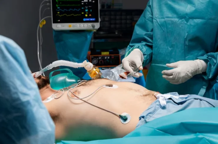
Thoracoscopy

Thoracoscopy is a minimally invasive diagnostic and therapeutic procedure used to examine the inside of the chest cavity (thoracic cavity). It involves inserting a small camera (thoracoscope) through tiny incisions in the chest to inspect the lungs, pleura (lining around the lungs), and other structures in the chest. Thoracoscopy is also known as “video-assisted thoracoscopic surgery” (VATS) when used for surgical purposes.
Thoracoscopy is performed under local or general anesthesia. A small incision is made in the chest, and a thin tube with a camera (thoracoscope) is inserted to visualize the lungs and pleural space. The procedure can be used for diagnostic purposes, such as identifying lung infections, cancers, and pleural diseases, or for treating conditions like pleural effusions (fluid buildup in the pleura).
Benefits of Thoracoscopy
- Minimally Invasive: Requires only small incisions, reducing pain, risk of complications, and recovery time compared to traditional open surgery.
- Quick Recovery: Most patients experience a quicker recovery and can return to their normal activities in a shorter time.
- Accurate Diagnosis: Provides clear visualization of lung and pleural conditions that might be difficult to diagnose through imaging alone.
- Therapeutic Uses: Besides diagnosis, thoracoscopy can be used for therapeutic interventions, such as draining fluid collections, performing biopsy procedures, and treating pleural diseases.
When is Thoracoscopy Needed?
Thoracoscopy is commonly used for:
- Pleural Effusions: Diagnosis and treatment of fluid buildup in the lungs.
- Lung Cancer: Determining the spread of cancer or obtaining biopsies.
- Infections: Diagnosing infections affecting the lungs or pleura.
- Benign Lung Conditions: Treating diseases like emphysema or fibrosis.
Thoracoscopy is a key procedure in modern pulmonology, offering both diagnostic clarity and therapeutic solutions with minimal patient discomfort.
